Trigonolampa miriceps Regan and Trewavas 1930
[Regan and Trewavas, Danish Dana expeds. 1920-1922, No. 6, 1930, p. 55, pl. 1, fig. 1.]
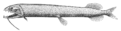
Descripton—
Trigonolampa resembles Stomias in general appearance, in the relative sizes and locations of the fins, and in having a long fleshy barbel on its chin. But it not only has a small light organ below the eye (as in Stomias), but also has a small luminescent patch close behind it, and likewise a larger triangular patch extending from close behind the eye back across the top of the cheek; these are its most distinctive characters. The one species of the genus yet known differs further both from Stomias (p. 147) and from Stomioides (p. 147) in a considerably deeper body (cf. fig. 67 with figs. 65, 66); also in that the tip of its lower jaw does not enclose the tip of its upper jaw when the mouth is closed; that the point of origin of its dorsal fin is in advance of its anal fin by a distance about as great as the diameter of the eye; and that it has only about 68 light organs in each of its ventral rows, as against 85 or 86 in Stomias (p. 147).
Color—
Not known, but probably black or very dark brown.[47]
Size—
The largest specimen yet seen (in the Museum of Comparative Zoology) is about 9 inches (230 mm.) long to the base of the caudal fin.
Range and occurrence in the Gulf of Maine—
Only three specimens have been seen yet. The first was taken in the eastern Atlantic by the Danish research vessel Thor in 1906 at a depth of about 600 fathoms; a second was found by Capt. John Toothaker in the stomach of a swordfish harpooned on the southern edge of Georges Bank in the summer of 1922,[48] and a third, now in the Museum of Comparative Zoology, was recorded simply as taken on Georges Bank about 1913. It reaches the slope of our outer Banks only as a stray from the mid-depths offshore.